Early detection is key to surviving melanoma, a type of malignant tumor responsible for more than 70% of skin-cancer-related deaths worldwide, but “suspicious pigmented skin lesions” (SPLs) are so common it’s impractical for doctors to check them all out. Now MIT researchers have developed a tool that can analyze skin photos taken with a smartphone to determine which SPLs should be evaluated by a dermatologist.
The researchers, who include professors Martha Gray, SM ’81, PhD ’86, James Collins, and Regina Barzilay and postdoc Luis Soenksen, PhD ’20, made use of deep convolutional neural networks, machine-learning algorithms often used to classify images.
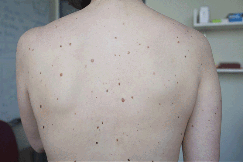
The team had dermatologists visually classify the lesions in 20,388 images from 133 patients at the Hospital Gregorio Marañón in Madrid and a number of publicly available images. The system was trained on 80% of those images and tested with the rest. It distinguished more than 90.3% of SPLs from nonsuspicious lesions, skin, and complex backgrounds. It also was able to classify the level of suspiciousness.
“Our research suggests that systems leveraging computer vision and deep neural networks, quantifying such common signs, can achieve comparable accuracy to expert dermatologists,” Soenksen says. The screenings could be done during routine primary care visits, or even by patients themselves.
Keep Reading
Most Popular
Ethically sourced “spare” human bodies could revolutionize medicine
Human “bodyoids” could reduce animal testing, improve drug development, and alleviate organ shortages.
Everyone in AI is talking about Manus. We put it to the test.
The new general AI agent from China had some system crashes and server overload—but it’s highly intuitive and shows real promise for the future of AI helpers.
Anthropic can now track the bizarre inner workings of a large language model
What the firm found challenges some basic assumptions about how this technology really works.
The foundations of America’s prosperity are being dismantled
Federal scientists warn that Americans could feel the effects of the new administration's devastating cuts for decades to come.
Stay connected
Get the latest updates from
MIT Technology Review
Discover special offers, top stories, upcoming events, and more.